
Navicular syndrome: everything you should know
Navicular inflammation, podotrochlosis, palmar foot pain, navicular bone disease: many names for a broad clinical picture
Navicular syndrome is a common lameness diagnosis in riding horses and often plunges horse owners into fear and anxiety, as it is still widely considered incurable. The good news is: that is not always the case. Over time, diagnostic tools have improved significantly and we have learned more about the disease and especially its causes, which is why today not all patients with navicular inflammation are automatically "hopeless cases".
Help, my horse has navicular!
To understand why navicular disease is so varied in prognosis and treatment, we need to look more closely at the anatomy. Because: every horse has a navicular! Strictly speaking, every hoof contains a so-called navicular complex. This consists of:
- navicular bone (a small elongated bone in the hoof that acts as a "deflection pulley" for the deep flexor tendon)
- ligaments of the navicular bone to the coffin bone and pastern bone (= upper and lower navicular bone ligaments)
- insertion of the deep flexor tendon, which runs over the navicular bone
- navicular bursa, which ensures smooth "rolling" of the tendon over the bone
- share of the coffin joint


All these components of the navicular complex can become diseased and cause the same symptoms. The symptoms are often diffuse and inconsistent: frequent stumbling, stiff gait, lameness especially on tight turns and on hard ground, avoidance of loading the rear part of the hoof (toe-first landing), often both forehooves affected. The same symptoms can however also occur when other structures in the back area of the hoof are painful. That is why in English-speaking countries the term "navicular syndrome" is often no longer used, but rather "palmar foot pain" or "caudal heel syndrome", which means something like "pain in the back area of the hoof / heel area syndrome".
Diagnosis
How can one then know exactly whether the navicular complex is really diseased or another structure? And if it is the navicular complex: which of its components?
The answer is often difficult in practice. By means of nerve block anaesthesia it can only be determined whether the back area of the hoof actually causes the pain, but not which part inside the hoof exactly. By means of X-ray only bones can be made visible, i.e. it can only be determined whether the navicular bone is healthy. In the past, the diagnosis of "navicular inflammation" was made as soon as enlarged vascular channels were visible in the navicular bone. These can however also have arisen from other problems (e.g. a previous, already healed coffin joint inflammation) and say nothing about the acute condition. Moreover, these navicular channels develop only over time and are therefore not yet visible on X-rays at the beginning of the disease.
Fortunately, another imaging procedure has spread in recent years also in the veterinary field, with which soft tissues can also be examined: magnetic resonance imaging (MRI). With this it is possible to make also tendons and ligaments visible. A reliable diagnosis of navicular inflammation can therefore only be made by means of MRI. In all other cases, it cannot be ruled out that another part of the hoof is actually causing the pain.
Causes of navicular inflammation
Since the navicular complex consists of so many different structures, there is also a whole range of causes that can irritate or damage each of these structures. In rare cases trauma can also lead to the disease; however, in the vast majority of cases it is overloading of the deep flexor tendon or too much pressure on the navicular complex, respectively reduced blood flow, which lead to navicular syndrome. Concrete causes of these problems can be:
- hoof stance:
- flat (negative) coffin bone angle: each 1 degree of angle reduction means 4% more load on the deep flexor tendon
- very steep hoof (reduced hoof mechanism)
- too long toe (additional load on the deep flexor tendon during break-over)
- too little "shock absorption" (missing soft tissue), e.g. due to contracted hooves or ossification of hoof cartilage
- too much weight for the hoof size (too small hooves or overweight)
- lack of movement
- unnatural movements (especially high speed on circular tracks, jumping, generally overloading of the forehand)
Is navicular syndrome treatable?
As mentioned at the beginning, navicular disease was long considered incurable, because one assumed a degenerative disease of the navicular bone. In current practice, however, it depends much more on which part of the navicular complex is exactly diseased (soft tissue or bone) and why (cause investigation), and in which stage the horse is. Bony changes are irreversible, but can often still be managed well. Problems on the involved tendons, bursa or ligaments are often protracted, but can depending on the cause often completely subside.
The treatment must therefore be individually tailored depending on the cause. Thorough cause investigation is important so that it can be addressed, e.g. via an optimised exercise load or changed husbandry conditions.
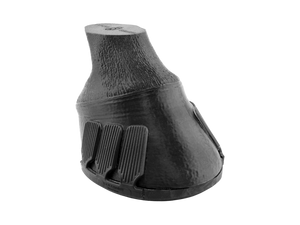
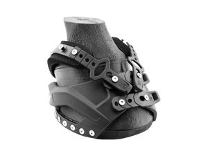
Since "true" navicular inflammation often cannot be distinguished from other pathological conditions in the back area of the hoof, the first measure should always also be the optimisation of hoof health. In some cases, correct shortening of the toe or elimination of contracted heels or a negative coffin bone angle is already enough to achieve an improvement. If necessary, it makes sense to apply an additional orthopaedic hoof protection (e.g. to elevate the heels, relieve the deep flexor tendon or for additional shock absorption).
Apart from that, painkillers, blood circulation-promoting medications or injections (into joint or bursa) may be indicated, depending on which structure is affected.
Sometimes one also hears that with a "navicular horse" a nerve resection ("neurectomy") is recommended or performed. In this procedure, the nerve responsible for stimulus transmission in the back area of the hoof is severed. As a result, the horse no longer feels this area and is therefore free of lameness. It must however not be forgotten that the actual cause persists and in most cases progresses unnoticed – the horse simply can no longer feel the worsening. Since the severed nerve strand is however responsible for the entire back area of the hoof, the horse also has no other perceptions any more in this area, e.g. of the nature of the ground. The horse is simply numb in the back area of the hoof, which sometimes means that other problems (e.g. abscesses or injuries) are overlooked.
In addition, it is not that uncommon that the nerve endings grow back together after a few years and the horse therefore regains its perception, with the lameness then often appearing significantly more severely (because the actual disease has progressed in the meantime).
A nerve resection must therefore be carefully considered and is not a simple solution, but rather a masking of the problem in cases where otherwise no pain-free life would be possible for the horse anymore.
Author: Nathalie Kurz
>> Sources
https://vetmed.illinois.edu/2020/09/11/navicular-syndrome/ https://extension.usu.edu/equine/research/equine-navicular-syndrome http://www.hufrollensyndrom.de/index.html https://www.pferdeklinik-aschheim.de/hufrollensyndrom-beim-pferd/ https://kernkompetenz-pferd.de/episode-23-hufrollen-erkrankung/ https://www.tierspital.uzh.ch/pferdekliniken/nervenschnitt/ https://www.usef.org/media/equestrian-weekly/is-it-navicular-syndrome-things-you-should-know